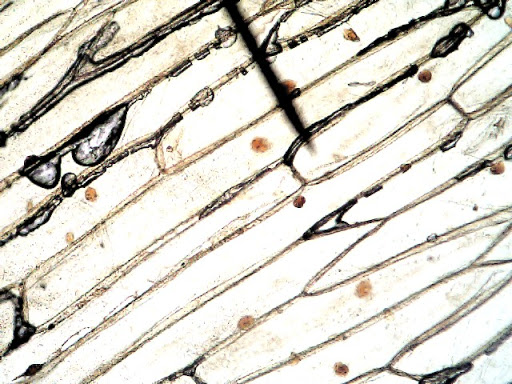
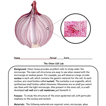
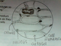
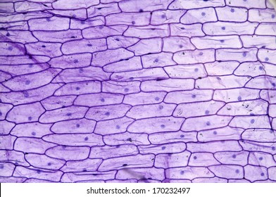
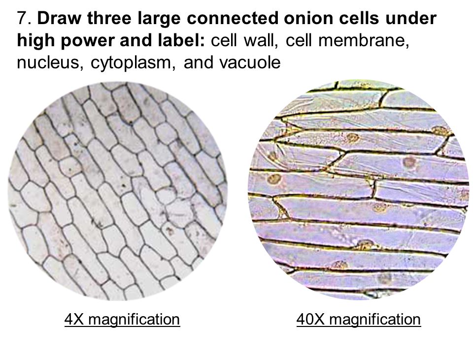
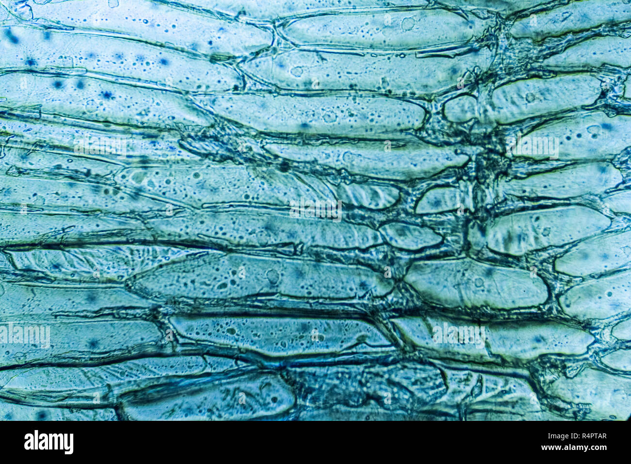
44 onion cells under microscope with labels
nfm.tobias-schaell.de › introduction-to-cellsIntroduction to cells worksheet pdf - nfm.tobias-schaell.de Jun 17, 2021 · Tell students to observe the PP slide with 3 images of cork cells under a microscope. Cells are the smallest living units within our body, but play a big role in making our body function properly. Many cells never have a large increase in size after they are first formed from a parental cell . yeson30.org › aboutAbout Our Coalition - Clean Air California About Our Coalition. Prop 30 is supported by a coalition including CalFire Firefighters, the American Lung Association, environmental organizations, electrical workers and businesses that want to improve California’s air quality by fighting and preventing wildfires and reducing air pollution from vehicles.
How to Observe Onion Cells under a Microscope - Blog, She Wrote Dec 19, 2015 ... Make a wet mount slide. Observe an onion cell under the microscope. Record our observations. Materials Needed for Observing Onion Cells. You ...
Onion cells under microscope with labels
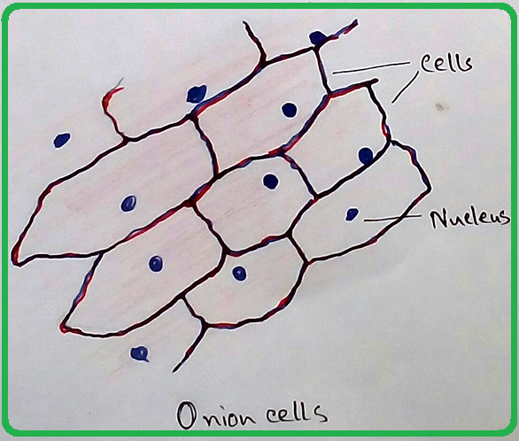
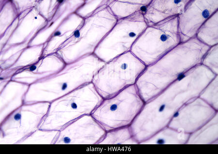
1,952 Onion Cell Images, Stock Photos & Vectors | Shutterstock Onion epidermis with large cells under light microscope. · Onion epidermis - microscopic view Stock Photo · High resolution light photomicrograph of Onion ... idoc.pub › documents › k-to-12-science-grade-7K To 12 Science Grade 7 Learners Material - Module How did the iodine solution affect the image of the onion cells? Q15. What parts of the onion cell can you identify? 8. Q16. Draw three to four onion cells as seen under the HPO. Label the parts you have identified. Indicate how much the cells are magnified. Of what importance is the contribution of the microscope in the study of cells? 85 Preparation and scientific drawing of a slide of onion cells including ... An onion is made up of swollen leaf bases separated by thin membranes of cells. In this exercise you will make a wet mount on a microscope slide, ...
Onion cells under microscope with labels. Onion cell preparation Set aside a clean microscope slide. 2. Carefully cut away a small, single layered ... This method allows students to view plant cells under the microscope. Epidermal onion cells under a microscope. Plant cells ... - Pinterest The Biology Lab Primer reiterates core information from lecture in a hands-on system focusing on the most ... Epidermal onion cells under a microscope. Onion Cells Under a Microscope - Requirements/Preparation ... An onion is made up of layers that are separated by a thin membrane. For this experiment, the thin membrane will be used to observe the onion cells. It can ... sciencequiz.net › newjcscience › jcbiologyThe Cell - ScienceQuiz.net The diagram shows a group of onion cells. The parts labelled A, B and C respectively are ... The diagram shows a plant cell as seen under a microscope. Two of the ...
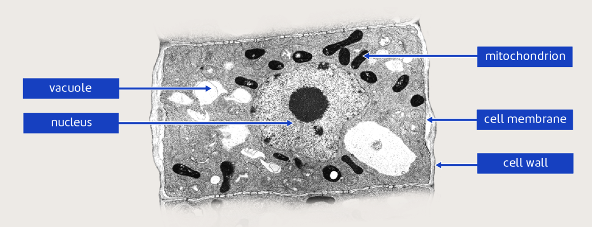
bmcbiol.biomedcentral.com › articles › 10Structure and function of mitochondrial membrane protein ... Oct 29, 2015 · Biological energy conversion in mitochondria is carried out by the membrane protein complexes of the respiratory chain and the mitochondrial ATP synthase in the inner membrane cristae. Recent advances in electron cryomicroscopy have made possible new insights into the structural and functional arrangement of these complexes in the membrane, and how they change with age. This review places ... Using a light microscope - Required Practical Review Biology Practical - Using a light microscope to observe, draw and label cells in an onion skin ... Using the microscope to look at animal and plant cells. School Science/How to prepare an onion cell slide - Wikibooks Tissue from an onion is a good first exercise in using the microscope and viewing plant cells. The cells are easily visible under a microscope and the ... Plant and Animal Cells Microscope Lab Students will observe onion cells under a microscope. Students will discover that their skin is ... Draw a diagram of one cheek cell and label the parts.
jnanobiotechnology.biomedcentral.com › articles › 10Nano based drug delivery systems: recent developments and ... Sep 19, 2018 · Nanomedicine and nano delivery systems are a relatively new but rapidly developing science where materials in the nanoscale range are employed to serve as means of diagnostic tools or to deliver therapeutic agents to specific targeted sites in a controlled manner. Nanotechnology offers multiple benefits in treating chronic human diseases by site-specific, and target-oriented delivery of ... ONION CELLS VIDEO - YouTube Sep 26, 2013 ... Video shows how to make a wet mount slide to view onion cells under the microscope. techcrunch.com › category › gadgetsGadgets • TechCrunch Nov 02, 2022 · Read the latest news, updates and reviews on the latest gadgets in tech. Coverage includes smartphones, wearables, laptops, drones and consumer electronics. Preparation and scientific drawing of a slide of onion cells including ... An onion is made up of swollen leaf bases separated by thin membranes of cells. In this exercise you will make a wet mount on a microscope slide, ...
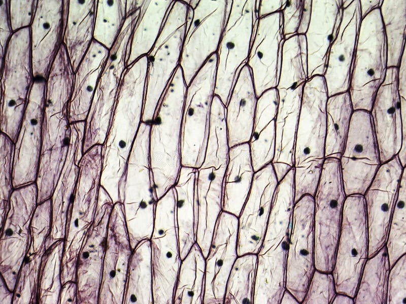
Biology Pictures: Onion Cells under Microscope
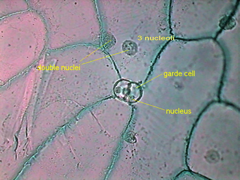
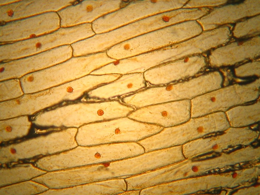
idoc.pub › documents › k-to-12-science-grade-7K To 12 Science Grade 7 Learners Material - Module How did the iodine solution affect the image of the onion cells? Q15. What parts of the onion cell can you identify? 8. Q16. Draw three to four onion cells as seen under the HPO. Label the parts you have identified. Indicate how much the cells are magnified. Of what importance is the contribution of the microscope in the study of cells? 85
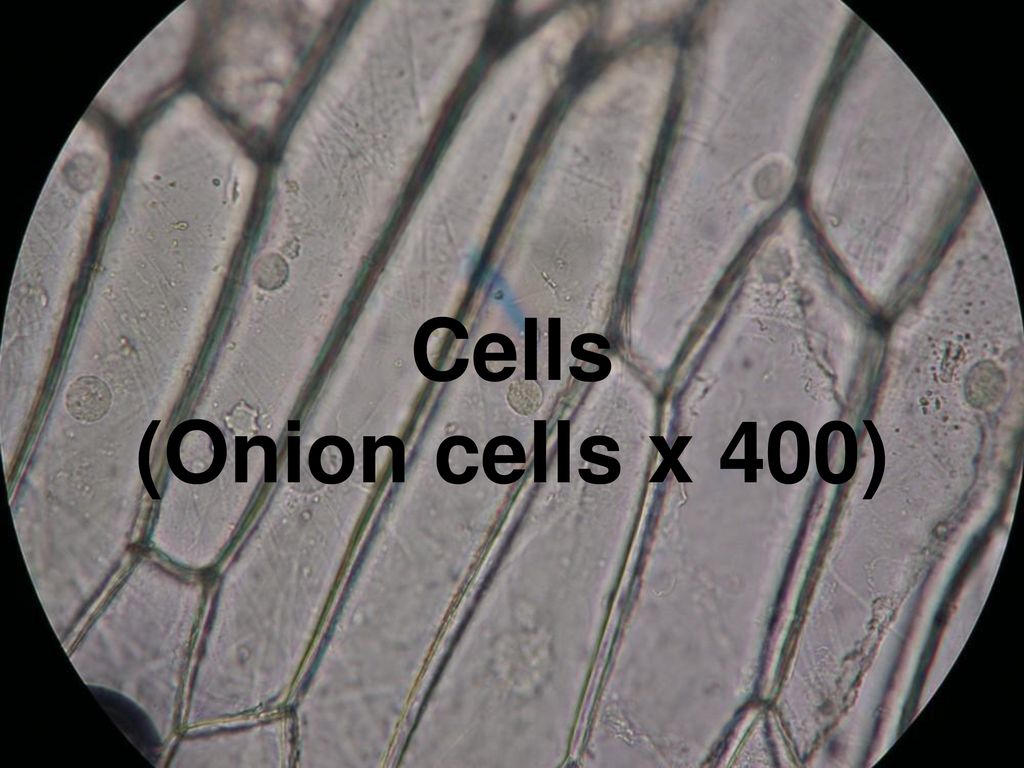
Cells (Onion cells x 400). - ppt download
1,952 Onion Cell Images, Stock Photos & Vectors | Shutterstock Onion epidermis with large cells under light microscope. · Onion epidermis - microscopic view Stock Photo · High resolution light photomicrograph of Onion ...
Mic-UK: How many onion skins are there?
3.3 Lesson: Cells under the microscope - Stile

Eisco™ Prepared Microscope Slide - Onion Mitosis, Logitudinal Section

Onion cells under a microscope hi-res stock photography and ...

Onion Cell Microscope Slide Experiment - YouTube
Onion Cell Microscope Lab
Intro to cells (article) | Khan Academy
What is the diagram of an onion cell? - Quora
Mic-UK: The inner epidermis of the onion bulb's cataphylls ...

Onion Cells at 400X Magnification

Cells Under A Microscope by Jaimarie Nelson
Personal Experience with Microscopes - AyushiSinhaMicroscopy

Onion skin cells under the microscope | iPad Case & Skin
167,915 Cell Wall Images, Stock Photos & Vectors | Shutterstock

Structure and Function of Cells (Learn) : Biology : Class 8 ...
Required Practical Review
Onion Peels Observed Under the Microscope | Confirmation Point
Onion cells light microscope hi-res stock photography and ...
File:Onion Cells.jpg - Wikimedia Commons

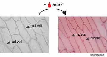
Onion cells fixed and stained with eosin

Cell structure Learning Intention: - ppt video online download
Mic-UK: MICROSCOPY UK / MICSCAPE - Onion epidermis, plasmolysis

Sketch the onion peel cell as seen under the microscope ...

File:Onion skin cells under a microscope.jpg - Wikimedia Commons
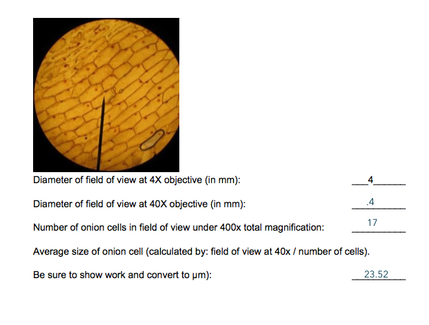
4 Diameter of field of view at 4x objective in mm): | Chegg.com
How do you observe the cells in onion peel under microscope ...
Onion cells under the microscope under two CONDITIONS ...
Photos Onion Cells Under Microscope That Stock Photo ...
Lab: Comparing Plant and Animal Cells - ppt video online download

Onion cells under microscopes | News | Wimbledon High School
321 Onion Cells Stock Photos - Free & Royalty-Free Stock ...

File:Onion Cells Under the Microscope.jpg - Wikimedia Commons

Iodine Stained Onion Cells Spiral Notebook by Ted Kinsman ...
What is the shape of an onion cell? Why? - Quora
Solved] draw and label three of the epidermal cells of Allium ...

Onion cell diagram Diagram | Quizlet
microbiology - What are the small white dots in an onion cell ...

Lab #1 microscope structure & function

Onion epidermis with large cells under microscope Canvas Print
Lesson 3: Onion Dissection & “Look at the Plant Cells” - Rs ...
Microscope view of onion cells Stock Photo - Alamy
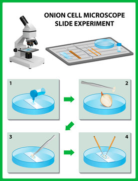
Microscopy. Onion Cell Microscope Slide Experiment. Vector ...
Post a Comment for "44 onion cells under microscope with labels"