39 diagram of the lungs with labels
Diagram Of The Respiratory System With Labels Stock Photos, Pictures ... Browse 157 diagram of the respiratory system with labels stock photos and images available, or start a new search to explore more stock photos and images. Newest results Human Lungs Diagram Cross section & anterior view of the human lungs. The respiratory system The human respiratory system medical illustration with internal organs Human Lungs Diagram Lungs Stock Illustrations - 2,509 Diagram Lungs ... - Dreamstime Download 2,509 Diagram Lungs Stock Illustrations, Vectors & Clipart for FREE or amazingly low rates! New users enjoy 60% OFF. 187,228,656 stock photos online. ... Labeled diagram with brain sections. Cranial nerves vector illustration. Labeled diagram with brain sections and its. Lungs. Respiratory organs detailed anatomy illustration on a ...
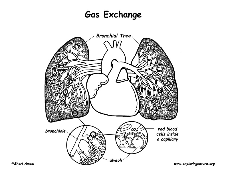
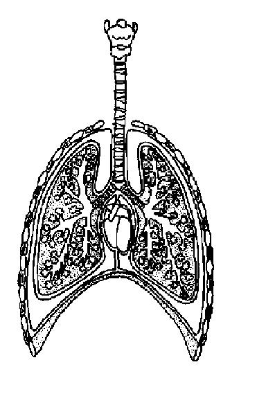
Label Lungs Diagram Printout - EnchantedLearning.com Read the definitions below, then label the lung anatomy diagram. bronchial tree - the system of airways within the lungs, which bring air from the trachea to the lung's tiny air sacs (alveoli). cardiac notch - the indentation in the left lung that provides room for the heart. diaphragm - a muscular membrane under the lungs.

Diagram of the lungs with labels
Lung Diagram | Free Lung Diagram Template - Edrawsoft The lung diagram template here clearly presents a pair of spongy on both side of the chest. Simply hitting on the template to learn more parts including pleura, ribs, bronchi, alveoli and more. Feel free to find out more human anatomy templates and symbols in the free download version. The Respiratory System (Label Diagram) - ScienceQuiz.net Match each pair by dragging from right to left. When complete click Check button. A Guide to Understand Lung with Diagrams | EdrawMax Online The lobes of the right lung are divided by fissures, namely oblique and horizontal fissures. An oblique fissure separates the lobes of the left lung. Surfaces (three): Lung has three surfaces, namely, costal, mediastinal, and diaphragmatic. These surfaces correspond to one particular thoracic area.
Diagram of the lungs with labels. Labeled diagram of the lungs/respiratory system. - SERC Jan 10, 2014 · View Original Image at Full Size. Labeled diagram of the lungs/respiratory system. Image 37789 is a 1125 by 1408 pixel PNG Uploaded: Jan10 14. Last Modified: 2014-01-10 12:15:34 Fully Labelled Diagram Alveolus Lungs Showing Stock ... - Shutterstock Stock Vector ID: 369984683 Fully labelled diagram of the alveolus in the lungs showing gaseous exchange. Vector Formats EPS 1114 × 800 pixels • 3.7 × 2.7 in • DPI 300 • JPG Vector Contributor S Steve Cymro Similar images See all Assets from the same collection See all Similar video clips The Lungs - Anatomy and Physiology - opentextbc.ca The lungs are pyramid-shaped, paired organs that are connected to the trachea by the right and left bronchi; on the inferior surface, the lungs are bordered by the diaphragm. The diaphragm is the flat, dome-shaped muscle located at the base of the lungs and thoracic cavity. The lungs are enclosed by the pleurae, which are attached to the ... Anatomy of the Lung | SEER Training Anatomy of the Lung. The lungs are the major organs of the respiratory system, and are divided into sections, or lobes.The right lung has three lobes and is slightly larger than the left lung, which has two lobes.. The lungs are separated by the mediastinum.This area contains the heart, trachea, esophagus, and many lymph nodes. The lungs are covered by a protective membrane known as the pleura ...
Human Lungs Diagram - Agaliprogram Given below is a labeled diagram of the human lungs followed by a brief account of the different parts of the lungs and their functions. Each lung reaches from the collarbone to the border between the chest and abdominal cavities. Source: en.wikipedia.org An illustration depicting cigarettes and a pair of lungs affected by smoking. Labeling Diagrams Of The Heart - Isacork Here is a heart labeling quiz for you. What is number eight pointing at in the heart diagram? This heart diagram for children is a detailed and accurate illustration of a human heart, with labels for each section. 1.Different Parts Of The Body ↓ 2.Major Veins ↓ 3.Right Atrium ↓ 4.Right Ventricle ↓ 5.Pulmonary Artery ↓ 6.Lungs Lungs Diagram Labeled Pictures, Images and Stock Photos Search from Lungs Diagram Labeled stock photos, pictures and royalty-free images from iStock. Find high-quality stock photos that you won't find anywhere else. Lungs label - Teaching resources 3728 results for 'lungs label'. Lungs Labelled diagram. by Rbowerkail. KS4 PE. The Lungs Labelled diagram. by Fayeroberts. KS4 Y10 Biology. Lungs Diagram Labelled diagram. by Jon9.
Label the lungs Vector Stock Images | Depositphotos 165 Label the lungs vector art & graphics are available under a royalty-free license. iv.pictured Set of 30 colorful vector icons of a medicine, health, corona virus and hygiene related objects. It represents a concept of medical protection, isolation, health safety and virus control equipment. DenisPotysiev Human anatomy organs. The Lungs - Labelled diagram Drag and drop the pins to their correct place on the image.. trachea, bronchi, bronchioles, Alveoli, Heart, Diaphragm , Rib, Intercostal Muscle . Anatomy and Physiology: The Lungs Diagram | Quizlet Anatomy and Physiology: The Lungs. The trachea divides into two bronchi. Provides air flow to and from the lungs. It also provides protection when a foreign object accidentally gets into the trachea, the ciliary cells get irritated and induce coughing to expel the object. The trachea also provides thermoregulation, humidify and warm the air ... Lungs: Anatomy, Function, and Treatment - Verywell Health The lungs are a major organ that is part of the respiratory system, taking in fresh air and getting rid of old, stale air. This mechanism of breathing also helps to allow you to talk. By taking in fresh air, the lungs are able to help oxygenate blood to be carried around your body. This is done by inhaling the air and bringing it in toward the ...

Early Anatomy Graphics - Diagram of Lungs - The Graphics Fairy
Labeled Diagram of the Human Lungs - Bodytomy Given below is a labeled diagram of the human lungs followed by a brief account of the different parts of the lungs and their functions. Each lung is enclosed inside a sac called pleura, which is a double-membrane structure formed by a smooth membrane called serous membrane.
Pulmonary Circulation - Through Heart and Lungs (Advanced*)
Lung Anatomy, Function, and Diagrams - Healthline Dec 14, 2018 · The lungs begin at the bottom of your trachea (windpipe). The trachea is a tube that carries the air in and out of your lungs. Each lung has a tube called a bronchus that connects to the trachea....

Respiratory System Diagram | Respiratory system anatomy, Respiratory ...
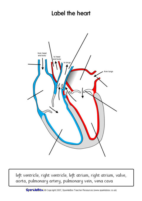
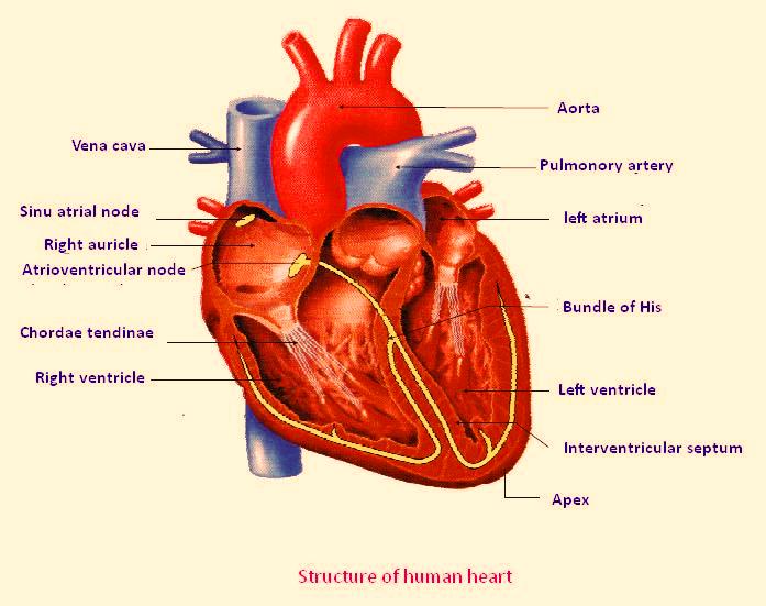
Label Lungs Diagram Printout - EnchantedLearning.com | Respiratory ... Diagram illustrating the paths of blood flow through the heart. Blood enters the heart through two large veins, the inferior and superior vena cava, emptying oxygen-poor blood from the body into the right atrium.
Related Items
Lobes of the Lung - SmartDraw Lobes of the Lung Lobes of the Lung Create healthcare diagrams like this example called Lobes of the Lung in minutes with SmartDraw. SmartDraw includes 1000s of professional healthcare and anatomy chart templates that you can modify and make your own. 4/22 EXAMPLES EDIT THIS EXAMPLE Text in this Example: Lobes of the Lung
Human Heart Diagrams
Label the Lungs Diagram | Quizlet ... superior lobe of right lung ... middle lobe of right lung ... inferior lobe of right lung ... superior lobe of left lung ... left main (primary) bronchus ... lobar (secondary) bronchus ... segmental (tertiary) bronchus ... inferior lobe of left lung ... Sets found in the same folder Bi 233: Labeling the Larynx 21 terms SunshineGirl79 the cell
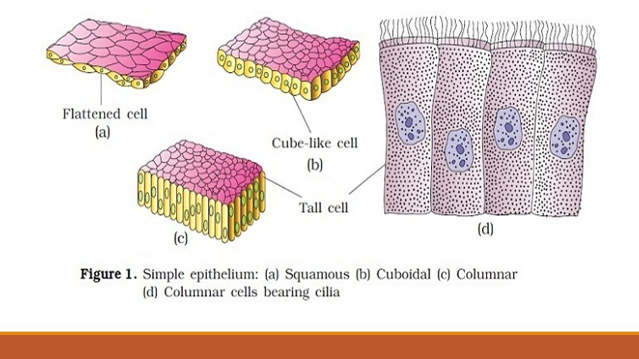
Types of epithelial tissue: simple, compound and specialized
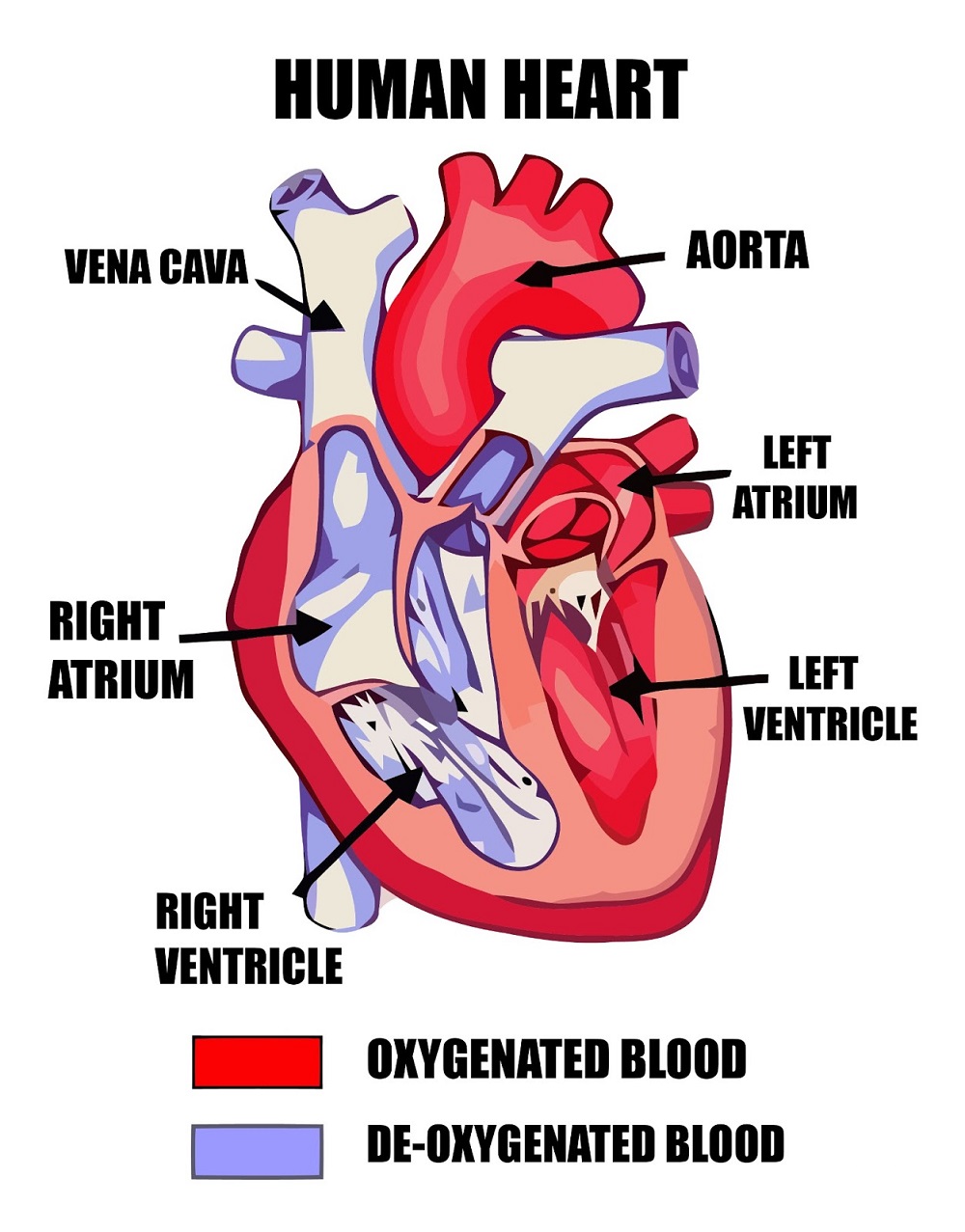
Diagram of Human Heart and Blood Circulation in It Four Chambers of the Heart and Blood Circulation. The shape of the human heart is like an upside-down pear, weighing between 7-15 ounces, and is little larger than the size of the fist. It is located between the lungs, in the middle of the chest, behind and slightly to the left of the breast bone. The heart, one of the most significant organs ...
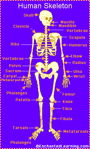
Human Skeleton - EnchantedLearning.com
Label Lungs Diagram Printout - EnchantedLearning.com Read the definitions below, then label the lung anatomy diagram. bronchial tree - the system of airways within the lungs, which bring air from the trachea to the lung's tiny air sacs (alveoli). cardiac notch - the indentation in the left lung that provides room for the heart. diaphragm - a muscular membrane under the lungs.
Free Unlabelled Diagram Of The Heart, Download Free Unlabelled Diagram ...
Lung Diagram Labelling Activity | Primary Resources | Twinkl This handy Lung Labelling Worksheet gives your children the opportunity to show how much they've learned about the human lung system. The beautifully hand-drawn illustration shows a lung diagram, labelled with blank spaces where learners can fill in its different components.
The Anatomy and Physiology of Animals/Respiratory System Worksheet ...
Lungs (Human Anatomy): Picture, Function, Definition, Conditions - WebMD The lungs are a pair of spongy, air-filled organs located on either side of the chest (thorax). The trachea (windpipe) conducts inhaled air into the lungs through its tubular branches, called...
Post a Comment for "39 diagram of the lungs with labels"